Anatomy Of Ribs Posterior : Posterior View of Rib Cage - Medical Illustration, Human ... - Skeletal system anatomy and physiology nurseslabs.. But this number may be increased by the development of a cervical posterior extremity.—the posterior or vertebral extremity presents for examination a head, neck, and tubercle. All 12 pairs of ribs attach to the building blocks of the spine (vertebrae) in the back. It branches from the ileocolic artery and may branch further to the appendicular artery. Head, neck, tubercle, and body of a rib. Includes images, video, and free quiz.
Be sure to subscribe to the visible body blog for more anatomy awesomeness! In the anatomical position, the scapula overlies the second to seventh ribs on the posterolateral aspect of the chest wall. The part of the muscle is thought to depress the ribs. Illustrations in anterior and posterior view of male torso and back, allowing the lines and regions used in surface anatomy to be displayed (midclavicular line, midline, pectoral region, sternal region.) ribs: Each rib articulates posteriorly with two thoracic vertebrae by the costovertebral joint.
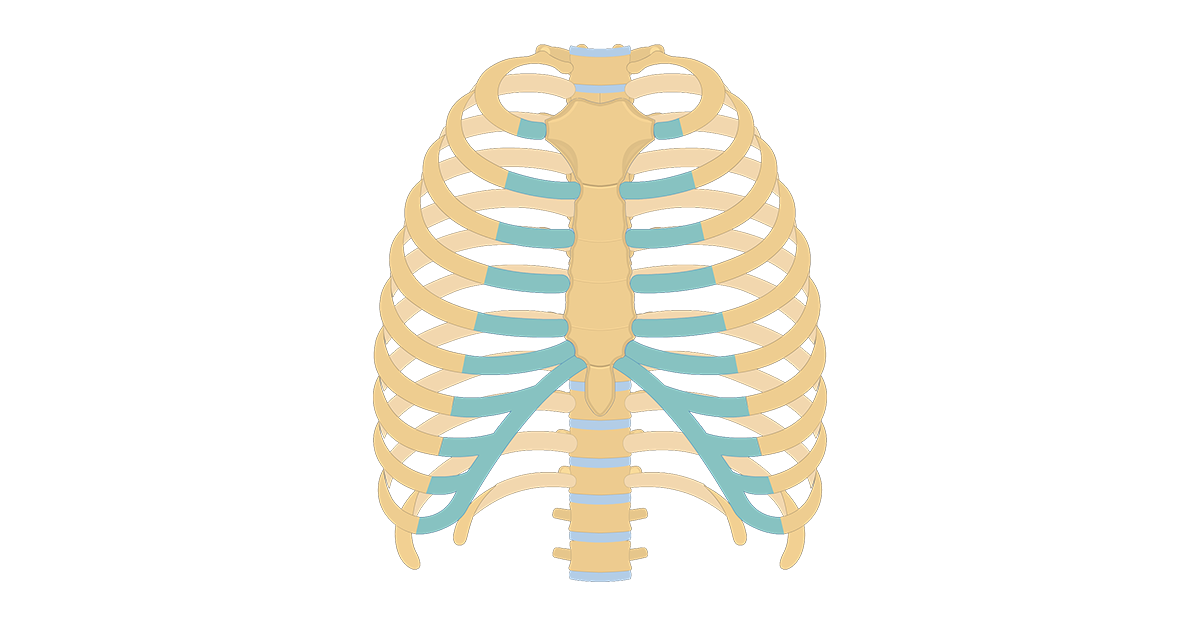
Structure of the Ribcage and Ribs from www.getbodysmart.com The ribs form the main structure of the thoracic cage protecting the thoracic organs, however their main function is to aid respiration3. Head, neck, tubercle, and body of a rib. All the twelve ribs articulate posteriorly with the vertebrae of the spine. Gross anatomy there are 12 pairs of ribs which are separated by intercostal spaces. It branches from the ileocolic artery and may branch further to the appendicular artery. 1.3 ribs anatomy and somatic dysfunctions. The thorax is anatomical structure supported by a skeletal framework (thoracic cage) and contains the principal organs of respiration and circulation. The posterior cecal artery is located in the abdomen near the lower intestines.
The number is the same in both males and females.
Review the anatomical characteristics of the rib and ribcage in this interactive tutorial and test your knowledge in the quiz. Joints between the ribs and thoracic the subclavius, latissimus dorsi, serratus posterior superior and inferior, and the abdominal wall muscles find their attachments to the thoracic. Represents the anatomy of the ribs and muscle attachments. In most tetrapods, ribs surround the chest, enabling the lungs to expand and thus facilitate breathing by expanding the chest cavity. Further details of its anatomical relations and muscle attachments can be found in its own section in this text. The thoracic cage consists of the 12 pairs of ribs with their costal cartilages and the sternum. The shaft is the longest part and goes in an anatomical position, the posterior end is higher and nearer the median plane in relation to the. Head of rib articulates with vertebra ribs move as a unit to accommodate breathing intercostal spaces = (spaces between ribs) • • •. Made up of thoracic vertebrae, ribs and… functions at upper end to connect the shoulder girdle and conn… The ribs form the main structure of the thoracic cage protecting the thoracic organs, however their main function is to aid respiration3. Major landmarks of a typical rib are the following: The nomenclature of the costal veins is the same as the arteries. They articulate with the vertebral column posteriorly, and terminate anteriorly as cartilage (known as costal cartilage).
This incision may be continued across the costal margin to open the abdominal cavity as in. Common characteristics of the ribs figs. It is split into ibrahim, af and darwish: The true ribs consist of 8 ribs, each on the left and right sides of the chest wall. Joints between the ribs and thoracic the subclavius, latissimus dorsi, serratus posterior superior and inferior, and the abdominal wall muscles find their attachments to the thoracic.
Learn Muscle Anatomy: Serratus Posterior Superior and Inferior from cdn2.hubspot.net The true ribs consist of 8 ribs, each on the left and right sides of the chest wall. The posterior cecal artery is located in the abdomen near the lower intestines. Posterior left rib fractures with injuries and nonunion of. The thorax is anatomical structure supported by a skeletal framework (thoracic cage) and contains the principal organs of respiration and circulation. Major landmarks of a typical rib are the following: Exposure of the posterior mediastinum is through the bed of the seventh or eighth ribs. Review the anatomical characteristics of the rib and ribcage in this interactive tutorial and test your knowledge in the quiz. These videos are for educational purpose only for the medical students like.
Head, neck, tubercle, and body of a rib.
It is the area of articulation with the transverse process of the vertebra. The subclavian artery and brachial plexus cross the rib posterior to anterior scalene muscle attachment and then run in contact with the bone on their way to the upper limb. Gross anatomy there are 12 pairs of ribs which are separated by intercostal spaces. In the anatomical position, the scapula overlies the second to seventh ribs on the posterolateral aspect of the chest wall. They are twelve in number on either side; These videos are for educational purpose only for the medical students like. Each pair articulates with a different thoracic vertebra on the posterior side of the body. Roughly speaking, this is the area of the chest. Skeletal system anatomy and physiology nurseslabs. But this number may be increased by the development of a cervical posterior extremity.—the posterior or vertebral extremity presents for examination a head, neck, and tubercle. In most tetrapods, ribs surround the chest, enabling the lungs to expand and thus facilitate breathing by expanding the chest cavity. Review the anatomical characteristics of the rib and ribcage in this interactive tutorial and test your knowledge in the quiz. It branches from the ileocolic artery and may branch further to the appendicular artery.
The subclavian artery and brachial plexus cross the rib posterior to anterior scalene muscle attachment and then run in contact with the bone on their way to the upper limb. Exposure of the posterior mediastinum is through the bed of the seventh or eighth ribs. 1.3 ribs anatomy and somatic dysfunctions. Skeletal system anatomy and physiology nurseslabs. Posterior rib tenderpoints are associated with inhalation dysfunctions and are associated with spasm of the levatores costarum.
Muscles of the Thoracic Wall - 3D Anatomy Tutorial - YouTube from i.ytimg.com They articulate with the vertebral column posteriorly, and terminate anteriorly as cartilage (known as costal cartilage). Common characteristics of the ribs figs. Posteriorly, the heads of the ribs interdigitate with the vertebrae and are numbered according to the inferior vertebra. This muscle is present posteriorly within the thoracic wall. The ribs are a set of twelve paired bones which form the protective 'cage' of the thorax. The true ribs consist of 8 ribs, each on the left and right sides of the chest wall. Be sure to subscribe to the visible body blog for more anatomy awesomeness! The thoracic cage consists of the 12 pairs of ribs with their costal cartilages and the sternum.
The number is the same in both males and females.
Gross anatomy there are 12 pairs of ribs which are separated by intercostal spaces. Ribs 3 to 9 are considered typical ribs. The lumbar plexus and its branches. by henry vandyke carter, henry gray (1918) anatomy of the human body. Exposure of the posterior mediastinum is through the bed of the seventh or eighth ribs. The nomenclature of the costal veins is the same as the arteries. Each rib articulates posteriorly with two thoracic vertebrae by the costovertebral joint. Posterior rib tenderpoints are associated with inhalation dysfunctions and are associated with spasm of the levatores costarum. Includes images, video, and free quiz. Made up of thoracic vertebrae, ribs and… functions at upper end to connect the shoulder girdle and conn… In the anatomical position, the scapula overlies the second to seventh ribs on the posterolateral aspect of the chest wall. This muscle is present posteriorly within the thoracic wall. Major landmarks of a typical rib are the following: Both muscles attach to various ribs and parts of the spine.
The subclavian artery and brachial plexus cross the rib posterior to anterior scalene muscle attachment and then run in contact with the bone on their way to the upper limb anatomy of ribs. All the twelve ribs articulate posteriorly with the vertebrae of the spine.


0 Komentar